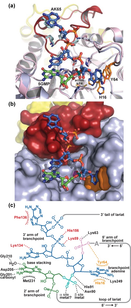
Figure 5.

Model of lariat RNA bound to Dbr1. (a) Superposition of the entire 5GMP•Dbr1 co-structure (pink) with the entire AK65•Dbr1(C14S) structure (light blue). The 5′ adenosine in AK65 (yellow in Figure 4b) has been omitted for clarity. The LRL and CTD elements are colored red and yellow, respectively, in both structures. The RMSD of all protein atoms is 0.18 Å. His16 is displaced from the active site in the 5GMP•Dbr1 structure, but engages in direct stacking interactions with the branchpoint adenine base in the AK65•Dbr1(C14S) structure. (b) The superposition of these two structures produces a model of the lariat branchpoint bound to Dbr1 (shown in the same orientation as panel a). Protein domains are colored as in Figure 2a. (c) Schematic of contacts between Dbr1 and the lariat branchpoint. Dashed lines denote hydrogen bonding contacts.
