
Figure 1.
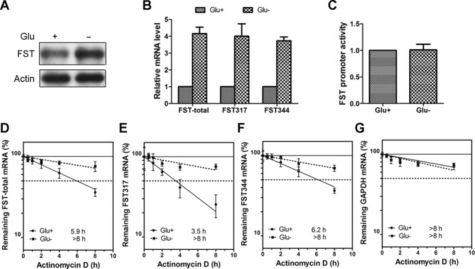
Glucose deprivation increases the stability of FST mRNA. (A) HeLa cells were incubated with (Glu+) or without (Glu−) glucose for 24 h and the protein level of FST was detected by immunoblotting. (B) HeLa cells were incubated with or without glucose and the mRNA levels of total FST, FST317 and FST344 were measured with real-time qPCR. Data shown are mean ± SD of three independent experiments. (C) HeLa cells transfected with FST promoter construct were incubated with or without glucose and luciferase activity was detected. Data shown are mean ± SD of three independent experiments. The mRNA levels of total FST (D), FST317 (E), FST344 (F) or GAPDH (G) in cells cultured with or without glucose were measured following treatment with actinomycin D for 0.5, 1, 2, 4 and 8 h. All the mRNA levels were normalized to 18S rRNA level. The half-life of each mRNA is defined as the time needed to reach 50% of its original abundance at time 0 h (dashed line). Data shown are mean ± SD of three independent experiments.