
Figure 5.
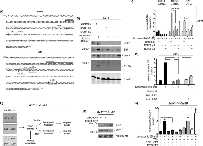
EGR1 contributes to bortezomib-induced apoptosis. (A) DNA sequences of the BS5 amplicon of human NOXA and BIM promoters. Primers are indicated by lines. Boxes show potential binding motifs for EGR1, Ets and MYC transcription factors. (B) DanG cells were transfected with control siRNA or two specific EGR1 siRNAs. After 66 h, cells were treated for 12 h as indicated. Western blot detected expression of EGR1, BIM and NOXA. Same extracts were transferred to two membranes and both were controlled by β-actin for equal loading. (C) DanG cells were treated as described in (B). Relative BIM, NOXA and EGR1 mRNA expression was determined by qPCR using PPIA mRNA as reference. Student's t-test *P < 0.05, n = 3. (D) DanG cells were transfected as indicated. Forty eight hours after the transfection, cells were treated with bortezomib for additional 24 h. Caspase activity was measured using caspase 3/7 activity assays. Student's t-test *P < 0.05, n = 3. (E) Working scheme: 3T9-MYClox/lox-CreER cells were transfected each with MYC-IRES-RFP, EGR1-IRES-GFP and combined. After 72 h, cells were FACS sorted. Cells were passaged and analyzed for expression of MYC and EGR1. Afterward, cells were treated with vehicle (EtOH) or 4-Hydroxytamoxifen (250 nM, TAM) for 72 h and subsequently treated with vehicle (DMSO) or bortezomib for additional 24 h and assayed for caspase 3/7 activity. (F) 3T9-MYClox/lox-CreER cells were transfected each with MYC-IRES-RFP, EGR1-IRES-GFP as outlined in (E). Western blot detected expression of EGR1, MYC and histone H3 (loading control). (G) 3T9-MYClox/lox-CreER cells were treated as described in (E) and relative caspase 3/7 activity was measured 24 h after bortezomib treatment (100 nM). One-way ANOVA *P < 0.05, n = 3.