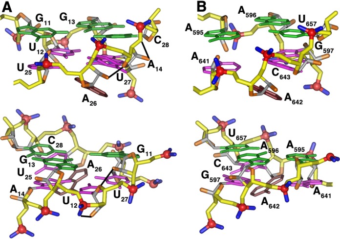
Figure 5.

Comparison of the non-canonical regions of (A) the aptamer RNA aptamer in complex with Bacillus r-protein S8 and (B) the spc mRNA in complex with E. coli r-protein. The structurally homologous base triples and adenine base are shown in green and brown, respectively. Intra-molecular hydrogen bonds unique to the aptamer (A) are depicted in black. The phosphate groups that interact with the S8 proteins have a very similar distribution (upper). The phosphate group of the additional residue in the core region of the aptamer is accommodated on the phosphate backbone strand distal to the protein surface (lower).
