
Figure 5.
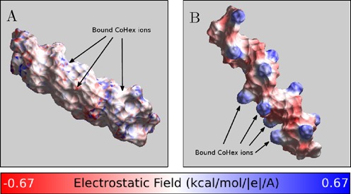
Charge neutralization patterns of NA duplexes by bound CoHex ions, assessed by the strength of the electric field near the NA-CoHex complex surface. (A) A-form mixed sequence RNA with CoHex counterions, which bind mostly in the major groove. (B) B-form mixed sequence DNA with CoHex ions, which are bound mostly externally. The specific snapshots are chosen to illustrate the internal (A) and external (B) binding modes from Figure 3 and reflect the actual average binding preferences; each snapshot has 15 bound (near neutralizing) CoHex ions, and is taken from the corresponding 320 ns-long all-atom MD simulation described in ‘Materials and Methods’. See Supplementary Data for a detailed visual characterization of CoHex ion distributions around these structures. The field is computed 3 Å away from the NA-CoHex complex molecular surface.