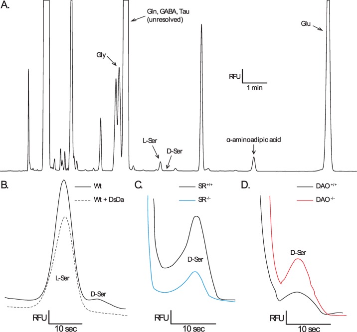
Figure 4.
Identification of capillary electrophoretic peaks from homogenized mouse retinas. (A) Electropherogram (relative fluorescent units (RFU) vs time) of an adult SR+/+ mouse retina showing the relative peak locations of NBD-F derivatized glycine (Gly), glutamine (Gln), γ-aminobutyric acid (GABA), taurine (Tau), l-serine (l-Ser), d-serine (d-Ser), α-aminoadipic acid (internal standard), and glutamate (Glu). (B) Normalized electropherograms of l-serine and d-serine in an adult SR+/+ vs the addition of d-serine deaminase (DsDa), which completely abolished the d-serine peak. (C) Normalized electropherograms of d-serine in an adult SR+/+ sample vs an adult SR–/– sample. (D) Normalized electropherograms of d-serine in an adult DAO+/+ sample vs an adult DAO–/–sample.

