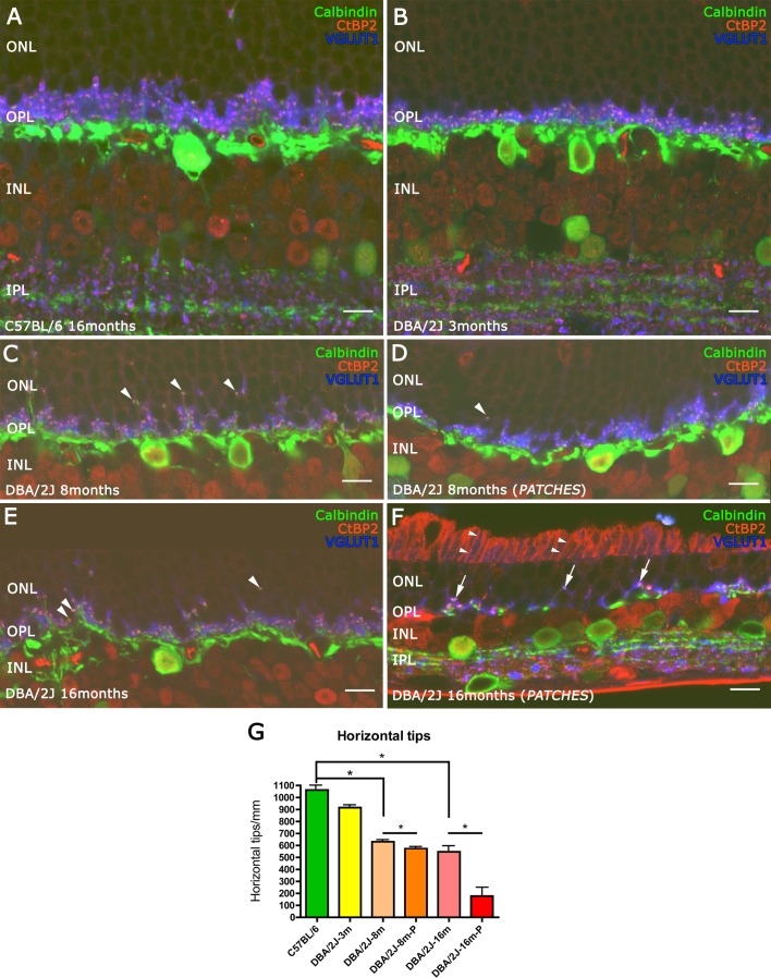
Figure 7.
Three specific markers of synaptic structure were used to study the connectivity between photoreceptor and horizontal cells. Antibodies against CtBP2 (red) and VGLUT1 (blue) were used to visualize the axon terminal structures of photoreceptor cells, and calbindin (green) was used to visualize horizontal cell dendrites. A thinning in the OPL was observed at 3 months old in the DBA/2J retinas (B) compared with C57BL/6 retinas (A). From 8 months (C, D) to 16 months (E, F), DBA/2J retinas showed growth of horizontal cells and synaptic contacts without VGLUT1 immunoreactivity (arrowheads). At 16 months old, inside the patches, only some synaptic contacts were complete ([F] arrows) and the plexus of the horizontal cells at OPL level were nearly absent. Quantification of horizontal cell terminal tips is shown in (G). *P < 0.05. Scale bars: 10 μm.

