
Figure 1.
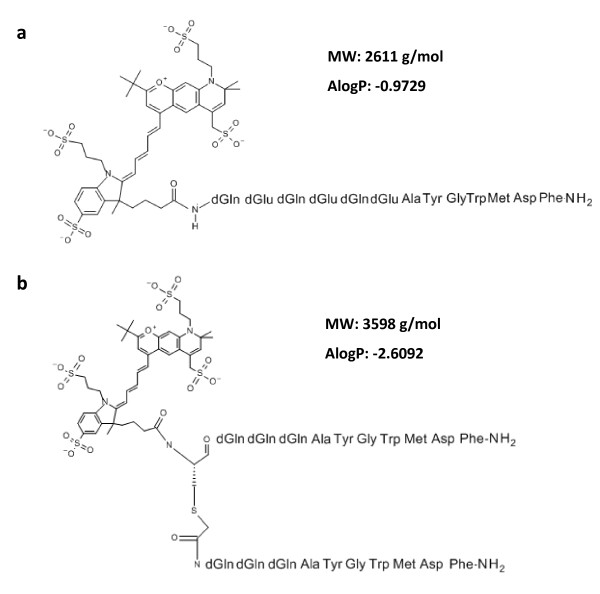
Structural formulas of optical CCKR binding peptide probes. (a) The probes differed in the spacer sequence connecting the NIR fluorochrome DY-754 and the CCK2R binding peptide. The probe QE displayed a spacer of three negatively charged d-glutamic acids alternating with three d-glutamines. (b) The probe bivQ had a bivalent nature, where each of the two minigastrin sequences was preceded by a spacer of three d-glutamines. Structural formulas were drawn with SymyxDraw.