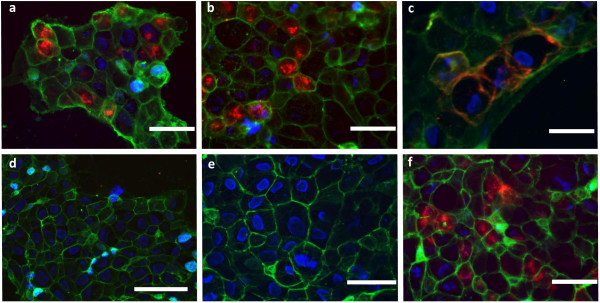
Figure 2.

The optical probes QE and bivQ showed specific binding and internalization in A431/CCK2R cells. We found intracellular uptake into A431/CCK2R cells at 37°C for the probes (a) QE and (b) bivQ and (c) cell membrane binding at 4°C (exemplarily for bivQ). (d) A431/WT cells showed no probe uptake at 37°C (exemplarily for QE). (e) Native A431/CCK2R showed no fluorescence in the NIR spectrum. (f) Similar internalization into A431/CCK2R cells was observed for the probe dQ-MG-754 [20] at 37°C. Displayed are representative fluorescence microscopy images of n = 3 experiments. Colour coding: red, DY-754 spectrum; green, cell membrane stain WGA-555; blue, cell nuclei stained with Hoechst 33258. Bars measure 100 μm.
