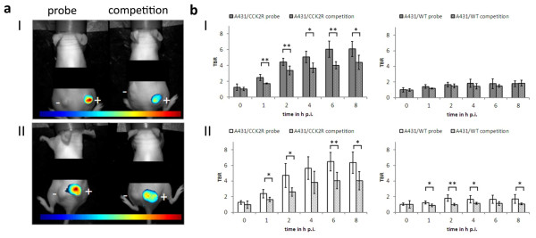
Figure 5.

Tumour accumulation of the probes QE and bivQ in vivo was highly specific. (a) Reduction of the fluorescence signals under competitive conditions for QE (I) and bivQ (II). (b) TBRs significantly decreased in A431/CCK2R tumours, but not in A431/WT tumours under competitive conditions for QE (I) and bivQ (II). *p < 0.05, **p < 0.01 (Student's t test). Means and standard deviations of n = 5 animals were calculated. Images were obtained with a NIRF small-animal scanner (excitation 615 to 665 nm, emission >750 nm), thresholded for their maximum fluorescence intensity (blue, lowest FI; red, highest FI) and overlaid with their respective white light image. Plus sign, A431/CCK2R tumour; minus sign, A431/WT tumour.
