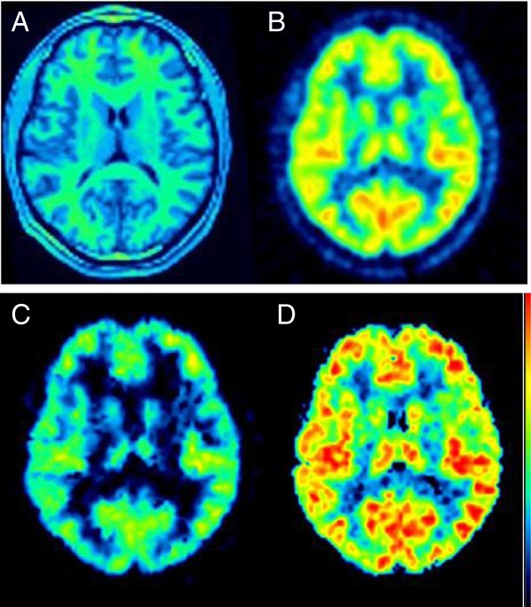
Figure 4.

Brain images. MRI (A), summed PET (B), and VT parametric images in a representative subject. The parametric images were calculated with the Logan plot (C) and spectral analysis (D) and are shown with the same color scale. Parametric images obtained with MA1 (not shown) were too noisy to be quantified.
