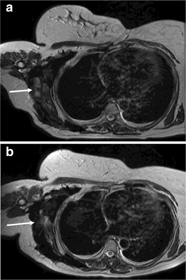
Figure 1.

Axial T2 weighted MRI images of the thorax at baseline (a) and following 10 months of pazopanib (b). A large plaque of fibromatosis encases the lateral right hemithorax extending into the axilla. Although the disease remained dimensionally stable the lateral component (arrows) demonstrated T2 signal drop from intermediate to low indicating a reduction in cellularity.
