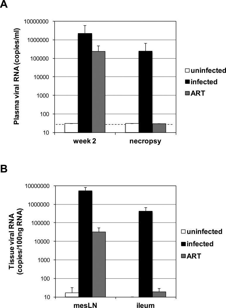
Figure 1.
(A) Plasma viral RNA detected at week 2 post-infection and necropsy from the uninfected animals, untreated RT-SHIV-infected animals, and ART-treated RT-SHIV-infected animals. The dotted line denotes the limit of detection of 30 viral RNA copies/ml. (B) Viral RNA detected in mesenteric LN and ileum samples of uninfected animals, untreated RT-SHIV-infected animals, and ART-treated RT-SHIV-infected animals is graphed per 100ng of total tissue RNA. Each bar represents the median of all samples in each group and error bars represent standard deviations.

