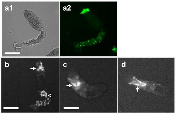
Fig. 3.
Higher magnification of cercariae exposed to human complement. Cercaria incubated with normal human serum for 2 h and stained with anti-neoC9 antibody (a1and a2) shows strong fluorescence in the anterior tip of the parasite (a2). Hoechst dye (b–d) strongly stains the tail when present (b, arrowhead) and the acetabular ducts (arrows in b, c, d). Scale bars are 100 μm in a 20× magnification field

