
Figure 2.
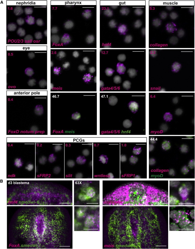
A Candidate Gene Approach Identified Tissue-Associated Transcription Factors Expressed in X1 Neoblasts from Regenerating Planarians
(A) FISH using previously known and new tissue-associated transcription factors with sorted X1 cells from prepharyngeal regions of amputated planarians. Percentages of X1 cells expressing the transcription factors are shown in the upper left corner. Total number of X1 cells counted: for protonephridia, a mixture of RNA probes POU2/3, odd-skipped related (osr), and sal-like (sall) n = 8/504; for the eye, ovo n = 1/194; for the anterior pole, a mixture of the RNA probes FoxD, prep, and notum n = 1/250; for pharynx, FoxA n = 54/704 and meis n = 20/777, with FoxA and meis coexpression observed in n = 7/15 of meis+ X1 cells; for the gut, hnf4 n = 337/1459 and gata4/5/6 n = 665/5249, with coexpression observed in n = 56/119 of hnf4+ X1 cells; for the muscle, collagen n = 72/1357 and the transcription factors myoD n = 47/732 and snail n = 45/1945, with myoD and collagen coexpression observed in n = 20/45 myoD+ X1 cells. Positional control genes were also expressed in wounded X1 cells: nou-darake (ndk, n = 23/6432), secreted related-frizzled 2 (sFRP-2, n = 9/4402), slit (n = 8/2763), wntless (n = 24/3367), and secreted related-frizzled 1 (sFRP-1; n = 7/732). Because some genes were expressed in multiple cell types, for some tissues, progenitor numbers will be an overestimate. DAPI labeled DNA (gray). Scale bars, 10 μm.
(B) Coexpression of the gut transcription factor hnf4, the muscle gene collagen, and the pharynx transcription factors FoxA and meis (magenta) with the neoblast marker smedwi-1 (green) in day 3 regenerating anterior blastemas or tail fragments (for the pharynx genes). Higher magnification on the right shows cells coexpressing both genes (scale bars, 10 μm). DAPI labeled DNA (gray). All images shown are maximal-intensity projections. Anterior, up. Scale bars, 100 μm.