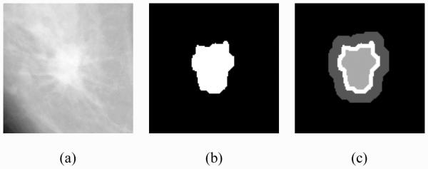
Fig. 5.

A (a) malignant mass, (b) its original segmentation mask and (c) different regions defined for computation of the contrast-based features: I1 (innermost lighter gray region of the mass), I2 (white region of the mass adjacent to its contour), and O (darker gray background region). Three sets of contrast features are computed from (1) between O and I whereby I is the interior segment of the mass within its contour (I1 + I2), (2) between O and I1, and (3) between O and I2
