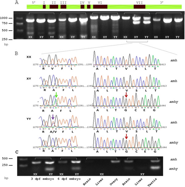
Figure 5.

Identification of Y-linked amh duplication. (A) Schematic illustration of the full length amh gene. Lines shaded with green, introns; Red boxes, exons; the Roman numerals outside the boxes indicate exon number. Four sets of PCR genomic fragments are presented under the respective parts of the gene for XX, XY and YY DNA samples. (B) DNA sequence traces of amh and amhy exon VII of three unrelated individuals: XX female, XY and YY males (GenBank: HG518783-7). Capital letters under the traces denote the deduced capable of encoding amino acids. Purple arrow, SNP in nucleotide position 1,274 of the full length amh gene in YY individual; green arrow, A > G substitution in nucleotide 1,275 of amhy between XY and YY individuals; red arrows, deletion starts in amhy from nucleotide position 1,403. (C) PCR for exon VII from cDNA of male and female 3 and 6 dpf embryos, brain, liver, and gonads.
