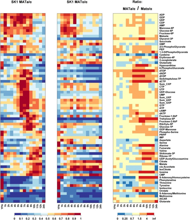
Figure 5.

Comparison of metabolite concentrations in sporulating SK1 MAT a/α cells and sporulation-deficient SK1 MAT α/α cells after transfer to sporulation medium. Metabolites were clustered according to similar temporal concentration profiles in the SK1 MAT a/α cells. Columns correspond to time points after transfer from YPA to sporulation medium. Each row corresponds to a metabolite concentration summarizing (left panel) the normalized concentration in sporulating cells, (middle panel) the normalized concentration in non-sporulating cells, and (right panel) the ratio of the concentrations in sporulating and non-sporulating cells. Concentrations were normalized to the maximum values observed over the whole time course. Data represent the average of two independent experiments. Concentrations are provided in Additional file 11. (Abbreviations: Sum_AXP = [ATP] + [ADP] + [AMP] likewise for Sum_UXP, Sum_GXP, and Sum_CXP; Rib/Xylu-5P = pool of ribulose-5P and xylulose-5P).
