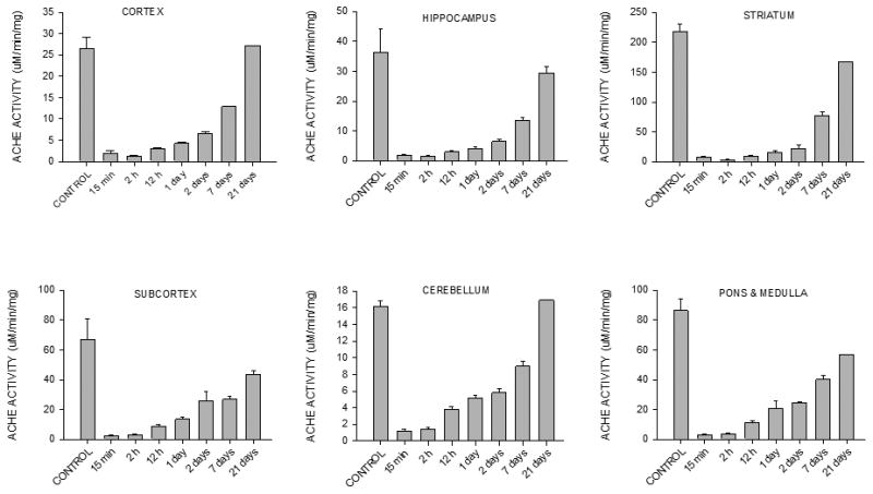
Figure 3. Experiment 1: Effect of DFP on brain AChE activity.

AChE activity was measured in six brain areas of rats treated with the pyridostigmine-ipratropium-DFP protocol (PI-DFP); pyridostigmine-ipratropium-water (PIW), or saline-saline-water (SSW) (see Fig. 2A). Enzyme activity was the same in the PIW and SSW groups, which were pooled together in the “CONTROL” group. The bars represent the mean ± SEM of 3-6 rats per time point, except at 21 days when there were only two rats, and therefore SEM was not calculated.
