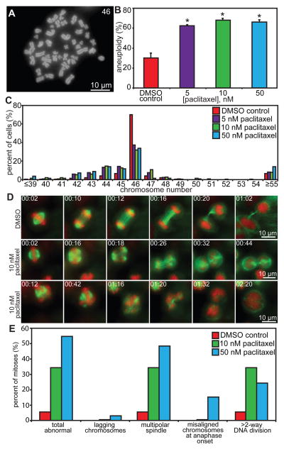
Figure 4. Low concentrations of paclitaxel increase aneuploidy and chromosome missegregation.
(A) Chromosome spread from a Cal51 cell containing 46 chromosomes. (B–C) Cal51 cells treated for 48 hours with 5, 10, or 50 nM paclitaxel show an increase in aneuploidy (B) that is predominantly near-diploid with little effect on triploidy or tetraploidy (C). n = 50 cells from each of 3 independent experiments. (D–E) Low dose paclitaxel increases abnormal mitotic events. (D) Cal51 cells expressing H2B-RFP (red) and GFP-tubulin (green) were filmed at 60× with 2-minute intervals during mitosis in DMSO (top row) or 10 nM paclitaxel (center and bottom rows). Shown are still frames from Videos S1–3. Time is shown in hours:minutes. Cells are able to divide in the presence of drug on multipolar spindles. (E) Quantitation of mitotic defects in Cal51 cells. The increase in defects is mainly due to multipolar spindles and >2–way DNA divisions. n=20–36 cells per condition. * = p < 0.05 as compared to DMSO control by Wilcoxon statistical analysis. Exact p values are listed in Table S2.

