
Fig. 6. Isolation of a small suppressive nanovesicle subpopulation by Antigen affinity chromatography.
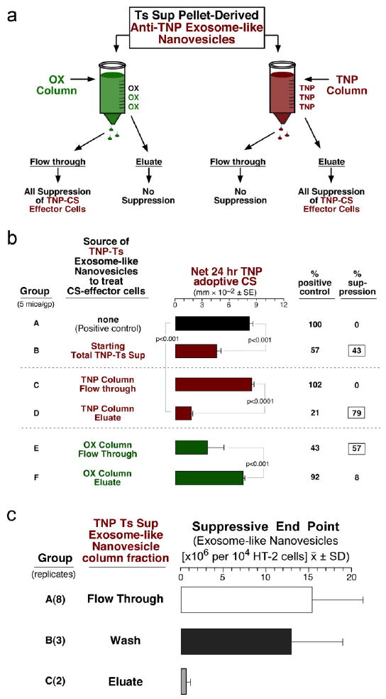
a. Suppressive TNP-Ts Sup nanovesicles were applied to either a column conjugated with TNP or OX. Only 12% of applied nanovesicles adhered to the TNP column and were eluted with dilute guanidine. b. TNP-Ts Sup vesicles from the TNP column flow through (FT) mediated no suppression (C), whereas the TNP-nanovesicles from the eluate had all the activity (D). The OX column FT, but not eluate had all the suppressive activity (E vs F). c. The eluate fraction from the TNP column strongly inhibited HT-2 cell viability (Group C), whereas the column wash (Group B), and the flow through (Group A) were not suppressive.