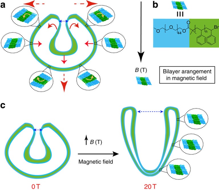
Figure 3. Mechanism of operation of the stomatocyte magneto-valve.
(a) Representation of the mechanism of magnetic deformation of the stomatocytes and the magnetic forces applied onto the membrane (red arrows). Yellow arrows show the net orientation of the individual polymer chains inside the bilayer to reach the preferred perpendicular orientation to the magnetic field. (b) Preferential perpendicular orientation of the bilayer in a magnetic field B and the chemical structure of the amphiphiles assembled into the bilayer. (c) High magnetic field deformation of the stomatocytes from spherical at 0 T into the prolate morphology at 20 T and the resulting increase in the size of the stomatocyte opening at high field (magneto-valve) as a result of the magnetic forces on the membrane.

