Figure 4.

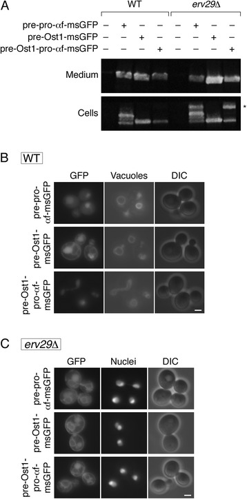
Effects on secreted msGFP constructs of including the α-factor pro region after the Ost1 signal sequence. (A) The analysis was performed as in Figure 1A, except that msGFP was fused to either the complete α-factor secretion signal (pre-pro-αf-msGFP), or the Ost1 signal sequence alone (pre-Ost1-msGFP), or the Ost1 signal sequence followed by the α-factor pro region (pre-Ost1-pro-αf-msGFP). The asterisk marks a band that may represent ER-localized msGFP molecules fused to the pro region. (B) The analysis was performed as in Figure 1B, except with the strains described in (A). Cells were stained with FM 4-64 to visualize the vacuolar membrane. Exposure times for the fluorescence images were 3oo msec. Scale bar, 2 μm. (C) The analysis was performed as in (B), except that vacuoles were not labeled, and the cells carried an erv29Δ allele and expressed nuclear-targeted DsRed-Express2. Exposure times for the fluorescence images were 300 msec. Scale bar, 2 μm.
