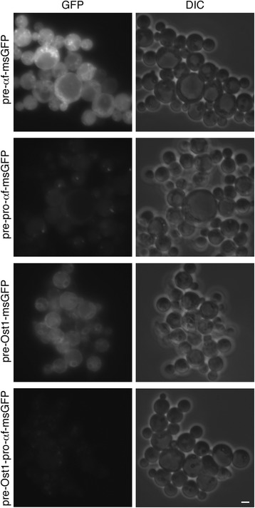
Figure 7.

Effects of the different signal sequences on intracellular accumulation of msGFP in P. pastoris . Strains of P. pastoris expressing the indicated msGFP constructs (see Figure 6) were imaged by fluorescence microscopy to detect GFP, and by differential interference contrast (DIC) microscopy to detect the cells. Representative groups of cells are shown. Exposure times for the fluorescence images were 200 msec. Scale bar, 2 μm.
