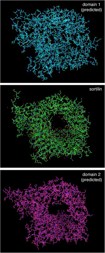
Figure 8.

Predicted structures of domains 1 and 2 of Vps10 compared to the known structure of sortilin. The protein sequences of S. cerevisiae Vps10 domain 1 (residues 22–737) and domain 2 (residues 719–1393) were submitted to the protein homology/analogy recognition engine Phyre (http://www.sbg.bio.ic.ac.uk/phyre/html/) [54], which detected the similarity to sortilin and generated PDB files for the predicted tertiary structures. A file for the experimentally determined structure of the human sortilin lumenal domain (PDB ID 3F6K) [53] was downloaded from the National Center for Biotechnology Information. To generate the images shown, these PDB files were opened with MacPyMOL using the default settings.
