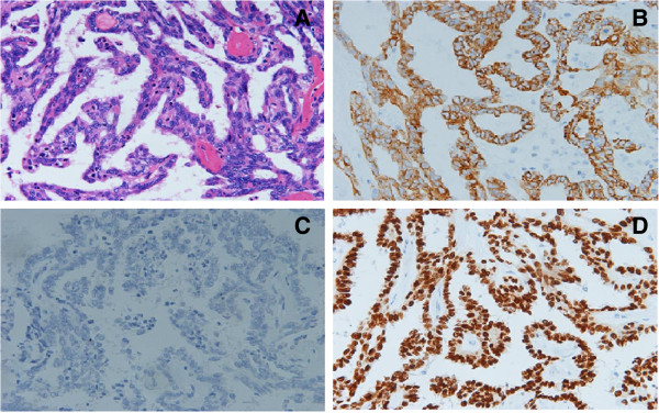
Figure 2.

Histopathological examination of thyroid showing papillary carcinoma. A: H & E stain showed moderately pleomorphic malignant oval to rounded epithelial cells. B, C, D: Immunohistochemical analysis revealed positive CK (B), TG (C) and TTF-1 (D) as markers of PTC. Magnification: ×200.
