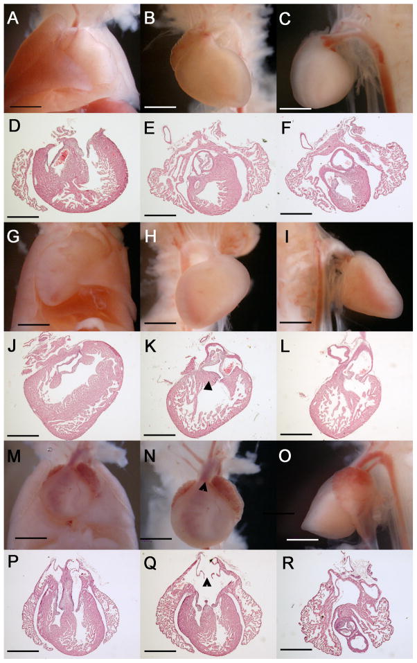
Figure 6.
Complex cardiovascular malformations in E17.5 Zic3 hypomorphs. Gross and histologic transverse sections from wild-type E17.5 embryos (A–F) and Zic3 hypomorphs (G–R). Wild type embryos have levocardial positioning in chest (frontal view, A) and proper orientation of chambers and great arteries (B, C frontal and lateral views, D–F transverse sections). A representative Zic3 hypomorphic embryo (G–L) demonsrates dextrocardial positioning in the chest (G) with right aortic arch (H, I frontal and lateral views) and DORV (J–L) with subaortic VSD (K, triangle). Another Zic3 hypomorphic embryo had mesocardial positioning in the chest (M), atrial isomerism, TGA, and an anteriorly-positioned aorta (N, triangle; O). Transverse sections demonstrate these defects as well as AVSD (P–R) and symmetrical venous valves (Q, triangle). All sections are in transverse plane, shown posterior to anterior. DORV, double-outlet right ventricle. VSD, ventricular septal defect. AVSD, atrioventricular septal defect. TGA, transposition of the great arteries. Scale bars, 2× magnification.

