
Fig. 5.
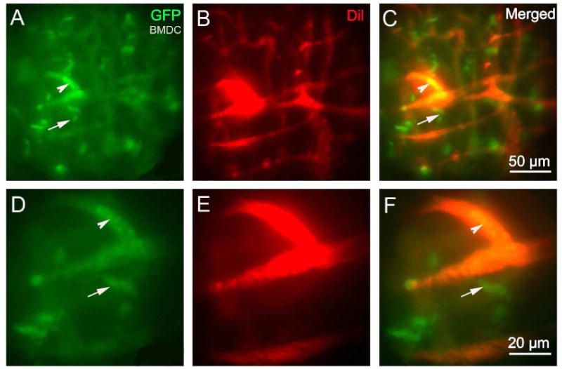
An open vessel-window preparation was used to visualize GFP+-BMDCs migration from cochlear lateral wall blood vessels in the lateral wall of a living mouse 18 months after bone marrow transplantation from a GFP donor mouse. (A) - (F) GFP+-BMDCs are shown migrated outside the blood vessel (red, labeled by Dil) under both low magnification (A) - (C) and high magnification (D) - (F). (Arrowheads point out GFP bone marrow cells that are inside of the blood vessels. Arrows point out GFP positive bone marrow cells that are outside of the blood vessels).