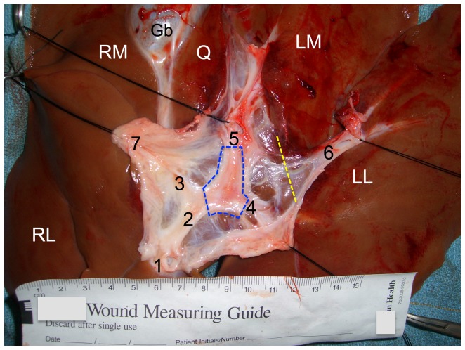
Figure 1. Dissection demonstrating the anatomy of the porcine intrahepatic portal venous system.
Ex vivo porcine liver, inferior aspect (scale in cm). The soft tissues overlying the portal venous system have been dissected and retracted with silk stay sutures. RL = right lateral lobe; RM = right medial lobe; LM = left medial lobe; LL = left lateral lobe; Q = quadrate lobe; Gb = gallbladder; 1 = cut orifice of main portal vein; 2 = intrahepatic portal vein; 3 = RM lobe portal vein branch; 4 = 1st LL lobe portal vein branch; 5 = cut orifice of 2nd LL lobe portal vein branch (proximal end); 6 = distal end of structure 5; 7 = pedicle containing the common bile duct and hepatic artery (reflected laterally by stitch). In this dissection the 2nd LL lobe portal vein branch was transected (the two ends are labeled as 5 and 6). The hepatic veins were not exposed in this dissection. The dashed blue polygon indicates the portion of the portal vein that was resected for the PVR injury mechanism. The dashed yellow line indicates where the cut was made across base of LL lobe for the LLLH injury mechanism. Scale = cm. [201 words].

