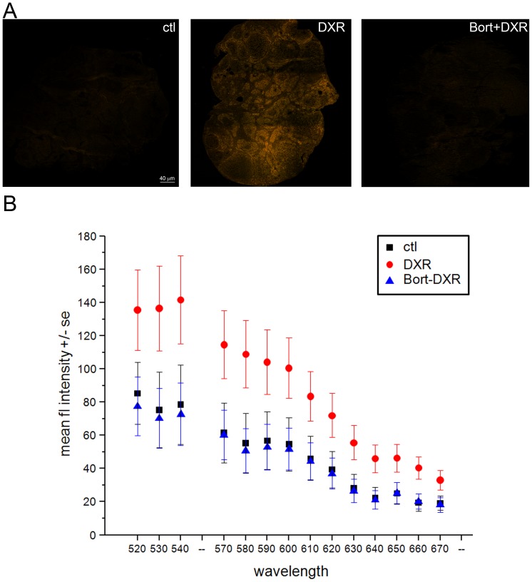
Figure 4. Bort pretreatment prevented DXR accumulation in the mouse ovary.
A. Micrographs of mouse ovarian sections obtained by spectral confocal imaging (ex. 488 nm, em. 520–720 nm). Images are overlays of all collected emission wavelengths. Bar = 40 µm. To ensure control autofluorescence was visible in print, images were adjusted equally to a threshold of 140 in Photoshop with no other image enhancements. B. Graph plots mean fluorescence intensity +/− SEM quantified from raw DXR fluorescence in ovarian sections representing the top, middle, and bottom third of the ovary. Emission profiles at 550–560 nm (cold finger) were not collected by the microscope to prevent direct detection of the excitation laser at that wavelength. Confocal parameters were identical from one sample to the next. DXR points are statistically significant from control and Bort-DXR with p<0.05, one-way ANOVA, Bonferroni means comparison.

