
Figure 3. Etoposide treatment does not directly decrease macrophage activation or the quality of antigen presentation by dendritic cells in LCMV-infected prf-/- mice.
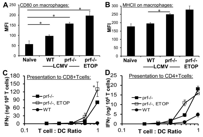
LCMV-infected prf-/- mice were treated with etoposide or carrier 5 days post-infection. LCMV-infected wild type mice treated with carrier are included for comparison. Surface phenotype of splenic macrophages (F4/80+) was assessed two days later by flow cytometry, including CD80 and MHC class II levels (A and B). Dendritic cells (CD11c+/ MHC II+) were magnetically sorted from collagenase treated spleens (pooled, 4 animals/group) seven days after infection and plated with LCMV-specific transgenic CD8+ or CD4+ T cells (P14 or SMARTA) to assess MHC class I and class II restricted presentation of endogenously acquired viral antigens. Antigen presentation to T cells was quantitated by IFN-γ production after overnight culture. No IFN-γ was detected when T cells of irrelevant specificity were cultured with DC’s (not shown). Data are the mean ± SE of 3-4 animals per group, representative of 3 independent experiments. *P<0.05