
Figure 5. Etoposide acts via selective destruction of activated effector T cells in LCMV-infected prf-/- mice.
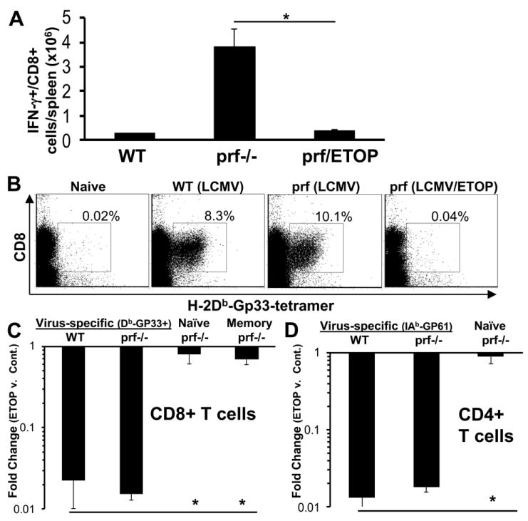
LCMV-infected WT or prf-/- mice were treated with etoposide or carrier 5 days after LCMV infection. Spleens were harvested 8 days post-infection after in vivo brefeldin administration for quantitation of T cells which were producing IFN-γ in vivo (A). In parallel experiments, day 8 spleen cells were stained using MHC-peptide tetramers (Gp33 in the context of Db) to delineate virus-specific CD8+ T cells. Representative plots of live gated-spleen cells from each group are shown. Percentage shown reflect % of live-gated/CD8+ cells. (B). Absolute numbers of virus-specific (Db-GP33 tetramer+), naïve (CD44lo), and quiescent memory CD8+ T cells were quantitated in LCMV-infected, etoposide (or carrier) treated animals (C). Fold change with etoposide treatment was calculated by comparing the total number of each cell population in spleens of carrier and drug treated animals. For assessment of quiescent memory T cells, ovalbumin-specific T cells primed by vaccinia-ova infection >1 month prior to LCMV infection were tracked. Virus-specific CD4+ T cells were enumerated with IAb-GP61-80 tetramer and compared to naïve CD4+ T cells in LCMV-infected, etoposide (or carrier) treated animals (D). *P<0.01 Data are ± SE, with >8 mice per group, from 3 or more experiments.