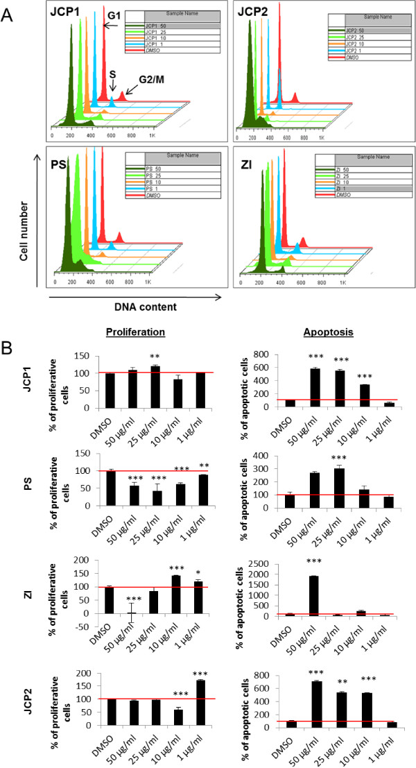
Figure 1.

Cell cycle analysis of MCF-7 cells. A: Histograms of the cell cycle distribution of MCF-7 cells after treatment with the control substance DMSO and the plant extracts JCP1, PS, ZI, JCP2 at concentrations of 1, 10, 25 and 50 μg/ml for 48 hours. G1, S and G2/M phases are marked with black arrows. Represented were the most prominent samples of 3–5 individual replicates. B: Calculation of proliferation and sub-G1 phase. Measurement of proliferation and apoptosis via cell cycle analysis after treatment with the vehicle DMSO (equates to 100%) and the plant extracts JCP1, PS, ZI, JCP2 at concentrations of 1, 10, 25 and 50 μg/ml for 48 h. As proliferative phases the sum of S and G2/M phases were calculated in percentages. As apoptotic fraction the sub G1-peak was measured. (mean ± SD, n = 5, ***P < 0.001, **P < 0.01, *P < 0.5, significantly different compared to control, unpaired t-test).
