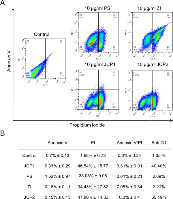
Figure 2.

Apoptosis determination via Annexin V/PI staining. A: Histogramms of Annexin V/PI- stained MCF-7 cells after treatment with 10 μg/ml of the four plant extracts in comparison with the control treatment for 48 h. Annexin V in conjunction with PI staining was used to distinguish early apoptotic (Annexin V- positive, PI negative; quadrant 1 of each panel) from late apoptotic or necrotic cells (Annexin V positive, PI positive; quadrant 2 of each panel). B: Table of quantitative analysis of single Annexin V or PI positive and double positive stained MCF-7 cells. Notably, sub-G1 phase positive cells from the cell cycle measurement were listed for comparison. Results are representative of three separate experiments.
