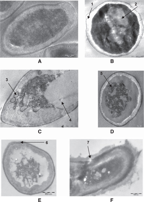
Figure 3.

Transmission electron micrograph of the 48 h grown C. albicans 04 cells in the absence and presence of sub-MICs of oils. (A) Untreated; intact cell wall, cell membranes and other organelles. (B, C) Cells grown in the presence of C. copticum at 90 μg.mL−1; loosening of cell membrane (1), disorganized cytoplasm (2), deposition of lipid globules (3), excessive vacuolization (4). (D,E) Cells grown in the presence of T. vulgaris at 90 μg.mL−1; disorganized protoplasm with receding of cell membrane (5), thickening of cell wall (6). (F) Cells grown in the presence of thymol at 90 μg.mL−1; lysis of cell wall and shrinkage of cell (7).
