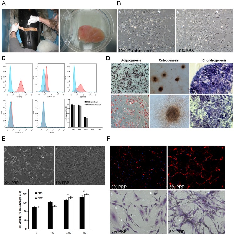
Figure 3. Dolphin PRP induces proliferation and phagocytic activity of dolphin ASCs.
(A) Adipose tissue was collected from the postnuchal fat pad from recent postmortem wild striped dolphins (Stenella coeruleoalba) (n = 2) and dolphin ASCs were derived and characterized. (B) Dolphin ASCs are plastic adherent and are able to be cultured in the presence of both 10% FBS and 10% dolphin serum. The morphology of ASCs treated with 10% dolphin serum appeared less elongated and senescent compared to those cultured with 10% FBS. (C) Dolphin ASCs were positive for mesenchymal cell markers CD90, CD44, and CD105 and were negative for hematopoietic cell markers CD34 and CD45. The histograms for CD90, CD44, and CD105 show the shift in the positive population in pink versus the non-stained sample in blue. CD34 and CD45 did not show positive reactivity, thereby confirming that the putative ASCs are indeed of mesenchymal origin. (D) Dolphin ASCs were capable of tri-lineage mesenchymal differentiation. ASCs were differentiated under standard in vitro conditions to adipocytes (Oil Red O staining), osteocytes (Alizarin Red staining) and chondrocytes (Alcian blue staining). (E) Dolphin ASCs treated in vitro with 2.5 or 5% dolphin PRP exhibited significantly increased proliferation, while those treated with 1% PRP were not different than controls. Proliferation rates in ASCs treated with the same concentrations of FBS were similar but significantly lower at 2.5 and 5% compared to PRP. Morphologically there was an increase in the number and density of ASCs cultured with 2.5 or 5% PRP compared to controls. Representative images of dolphin ASCs treated with 0 or 5% dolphin PRP are shown. Results of MTS assays are mean ± SD of colorimetric signal from treated cells in the presence of 1, 2.5, or 5% PRP compared to the absence of PRP. Every condition was assayed in quadruplicate in three different experiments for both lines of dolphin ASCs. (F) In addition to inducing proliferation of ASCs, treatment with 5% PRP stimulates phagocytic activity in dolphin ASCs. Red fluorescent microspheres were highly phagocytosed by ASCs in the presence of PRP compared to those without PRP (upper panels). Similarly, when fixed and stained with Giemsa, there were clearly more ASCs indicating increased proliferation. Also visible are the increased number of microspheres which have been phagocytosed within the ASCs treated with 5% PRP compared to fewer microspheres inside untreated ASCs. Asterisks denote significant difference compared to controls (0% or PRP or FBS) * P<0.05.

