
Figure 2.
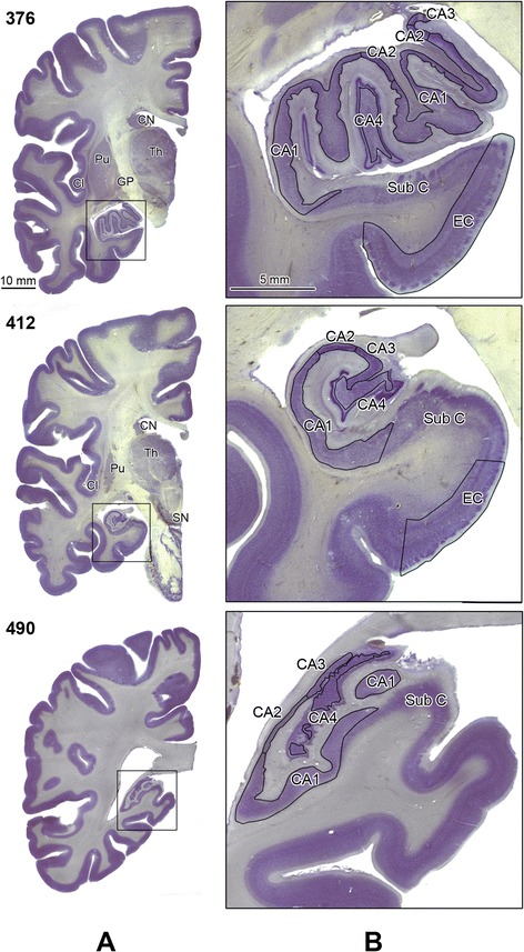
Delination of the Ammons horn sectors and the posterior portion of the entorhinal cortex. Panel A shows CV-stained hemispheric sections on the level of the hippocampal head (s. 376), body (s. 412) and tail (s. 490). The rectangle in panel A marks an area that was enlarged 5-fold and is shown in panel B. Stereologically examined structures are labeled as: sectors CA1, 2, 3, 4; EC, entorhinal cortex; CN, caudate nucleus; Pu, putamen; GP, globus pallidus; Cl, claustrum; Th, thalamus; and SN, substantia nigra. The subicular complex is labeled as Sub C. Calibration bar length: 10 mm and 5 mm.