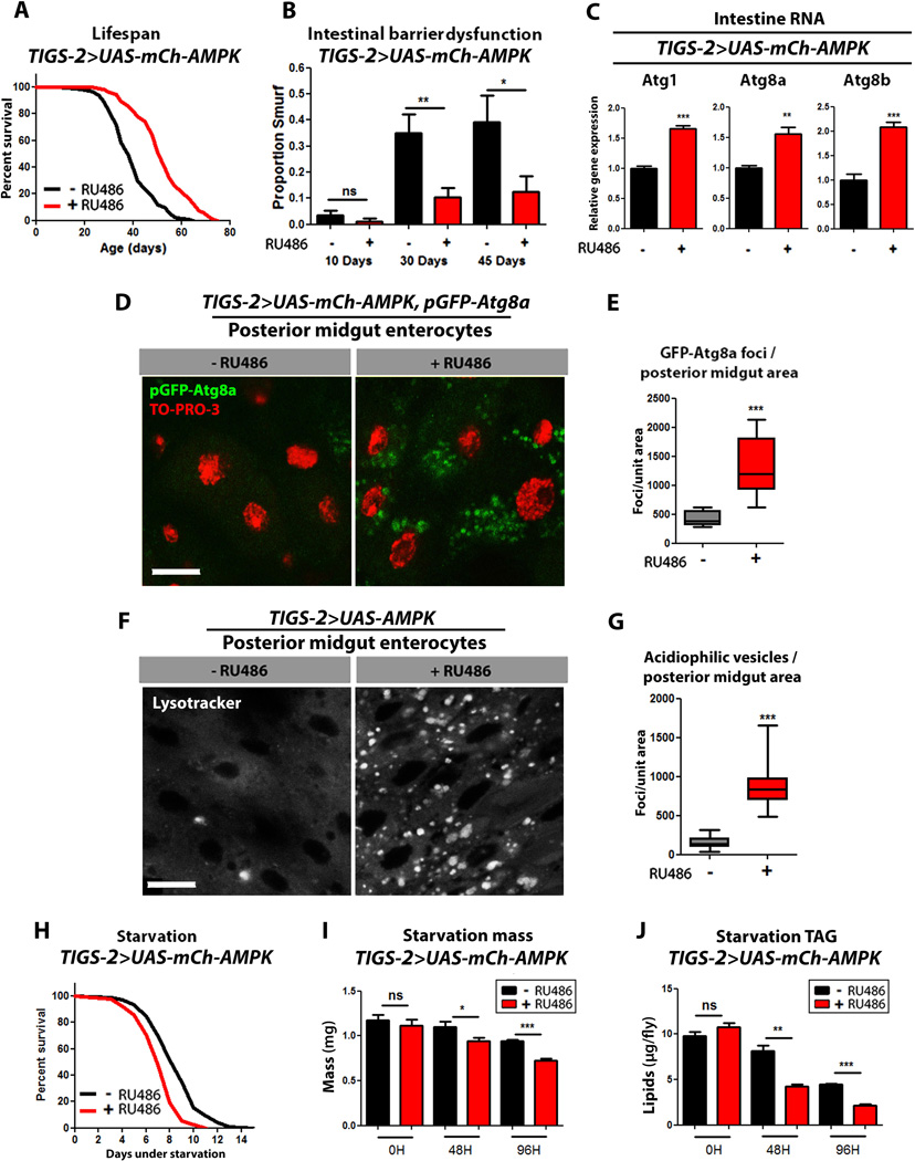
Figure 5. Intestinal AMPK activation maintains intestinal homeostasis during aging and extends lifespan.
(A) Survival curves of TIGS-2>UAS-mCh-AMPK females with or without RU486-mediated transgene induction (p<0.0001; log-rank test; n>116 flies).
(B) Intestinal integrity during aging in TIGS-2>UAS-mCh-AMPK females with or without RU486-mediated transgene induction (p<0.01, at 30 days, p<0.05 at 45 days; binomial test; n>127 flies/condition).
(C) Expression levels of autophagy genes in from intestines of TIGS-2>UAS-mCh-AMPK female flies at 10 days of adulthood with or without RU486-mediated transgene induction. (t-test; n>3 of RNA extracted from 15 intestines/replicate).
(D) GFP-Atg8a staining. Representative images of enterocytes from the posterior midgut of 10 day old TIGS-2>UAS-mCh-AMPK, pGFP-Atg8a females with or without RU486-mediated transgene expression (red channel-TO-PRO-3 DNA stain, green channel-GFP-Atg8a, scale bar represents 10µm).
(E) Quantification of posterior midgut GFP-Atg8a foci (p<0.0001; t-test; n>10 confocal stacks from posterior midgut/condition; one fly per replicate stack).
(F) Lysotracker Red staining. Representative images of posterior midgut enterocytes from 10 day old TIGS-2>UAS-AMPK females with or without RU486-mediated transgene induction (scale bar represents 10µm).
(G) Quantification of acidophilic vesicles (p<0.0001; t-test; n>10 confocal stacks from posterior midgut/condition; one fly per replicate stack).
(H) Survival curves without food of TIGS-2>UAS-mCh-AMPK females with or without RU486-mediated transgene induction (p<0.001; log-rank; n>257 flies).
(I) Body mass during starvation of TIGS-2>UAS-mCh-AMPK females with or without RU486-mediated transgene induction (p<0.05, at 48hours and p<0.01 at 96 hours of starvation; t-test; n>6 samples/condition; 10 flies weighed/sample).
(J) Whole body lipid stores during starvation of TIGS-2>UAS-mCh-AMPK females with or without RU486-mediated transgene induction (p<0.01 at 48 hours, and p<0.001 at 96 hours of starvation; t-test; n>3 samples/condition/timepoint; lipids extracted from 5 flies/sample).
Data are represented as mean ± SEM. RU486 was provided in the media after eclosion at a concentration of 25µg/ml (E, F) and 100µg/ml for all other figures.

