
Figure 1. Examples of 99mTc-MAA Scans during Pre-SIRT Evaluation.
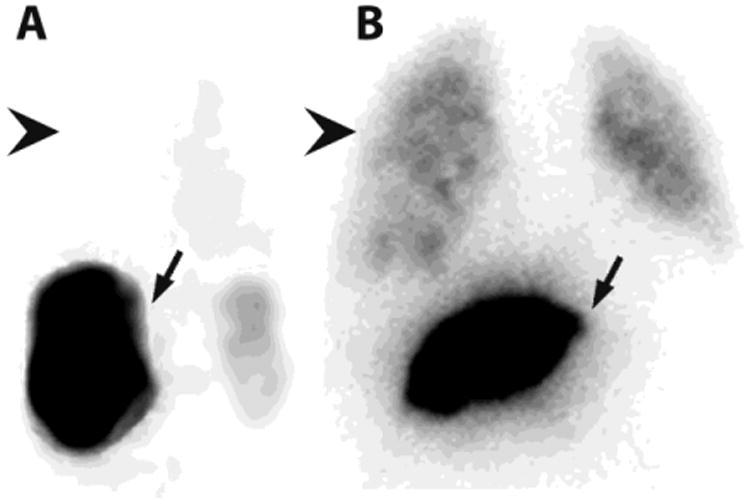
After 99mTc-MAA is injected into the hepatic artery to be treated during SIRT, the patient is transferred to nuclear medicine for SPECT and planar imaging to calculate the lung shunt fraction (LSF). A. In patients with little intratumoral vascular shunting (4%), activity predominates in the liver (arrow), with little or no activity in the lung (arrowhead). B. In patients with a great deal of shunting (12%), lung activity (arrowhead) is quantified and compared to liver activity (arrow) to calculate the LSF, which will determine the safe dose of radioembolization particles that may be administered during SIRT.