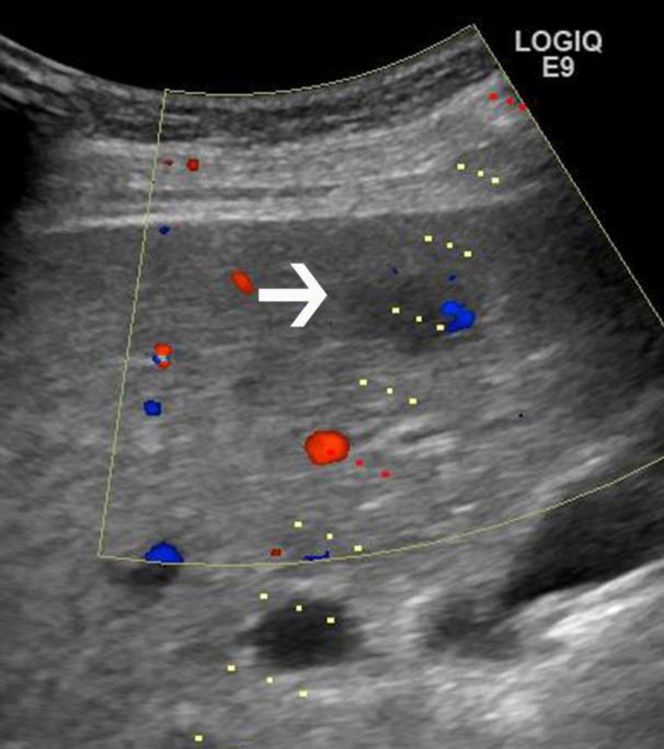
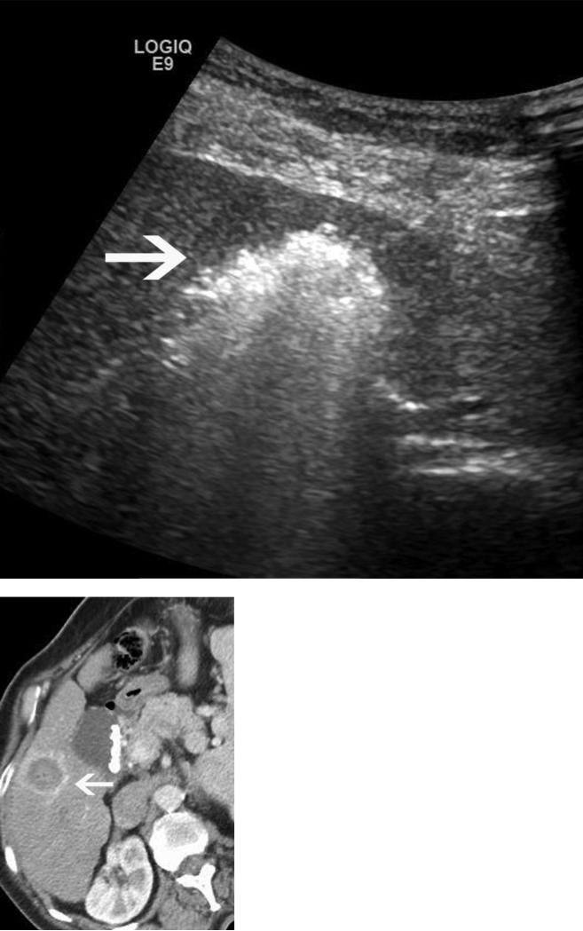
Figure 5.
Microwave ablation performed in a cirrhotic patient with an 18 x 15mm HCC. The preablation MRI demonstrates arterial enhancement (a) and washout (b) within the tumor (arrows), consistent with HCC. Intraprocedural US (c) redemonstrates the hypoechoic lesion (arrow), with the steam cloud encompassing the lesion in (d). A single antenna was run for 5 minutes at 65W, creating a spherical ablation zone measuring 2.8 × 2.8 cm, shown on axial post procedure contrast enhanced CT (arrow, e,).


