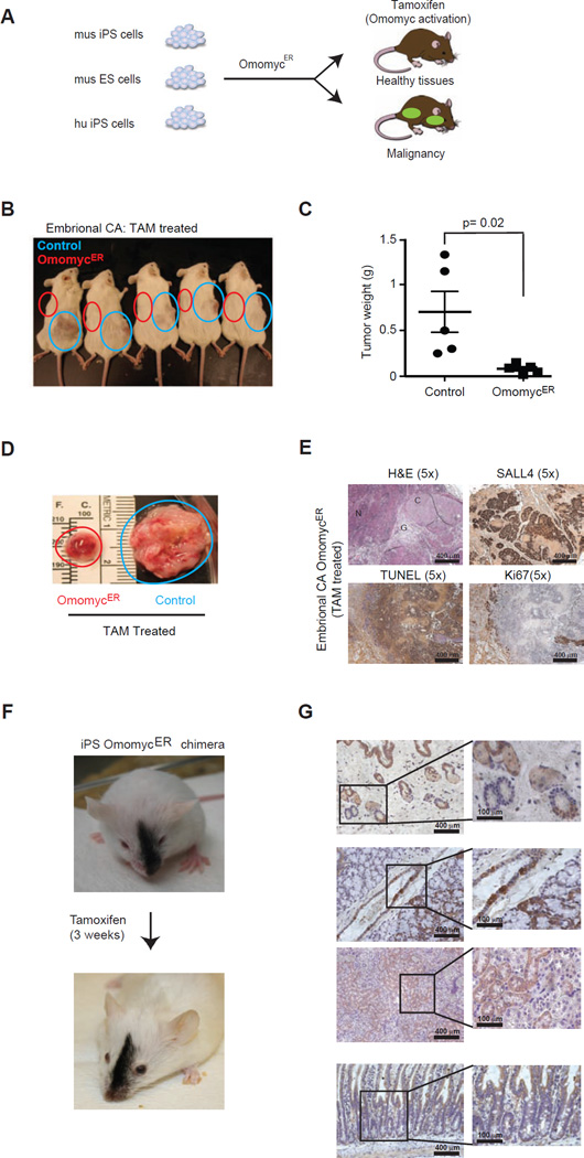
Figure 1. Aggressive embryonal carcinomas are sensitive to OmomycER treatment.
A, Schematic of mouse iPS (mu iPS), ES (mu ES) and human iPS (hu iPS) engineer with and without inducible dominant negative MYC allele OmomycER. B, Animals bearing embryonal carcinomas derived either from iPS cells expressing OmomycER (red circle) or control vector (blue circle) and treated with tamoxifen (TAM). C and D, Comparison of tumor weights following TAM treatment. The blue and red circles highlight the position (b) and the size (c) of the xenografted tumors. E, Histopathology of primitive embryonal carcinomas (Embryonal CA) stained as indicated; F, Representative chimeric animal before and after a 3 week course of tamoxifen (TAM) treatment to activate the dominant negative MYC construct in iPS cell derived tissues; (the same schedule was used in tumor treatment studies); G, Immunohistochemical stain for citrine identifies tissues derived from OmomycER expressing iPS cells in different organ sites (panels from top): hair follicles, upper gastrointestinal tract and adjacent thyroid gland, kidney, lower gastrointestinal tract and mucosal villi.

