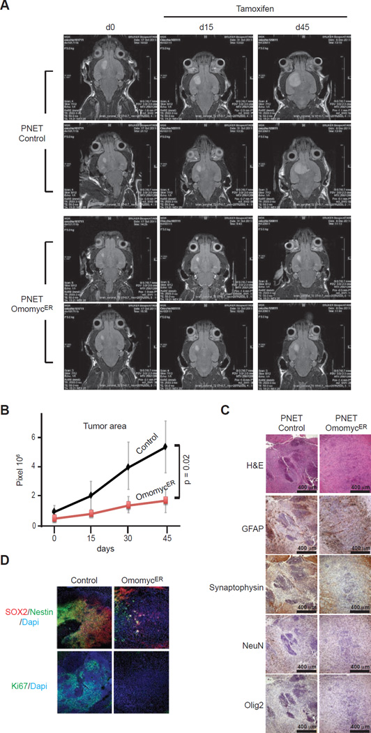
Figure 2. Primitive neuroectodermal tumors (PNETs) derived from human iPS cells respond to OmomycER activation in vivo.
A, Magnetic resonance imaging (MRI) of vector control (Control) and OmomycER expressing PNETs before tamoxifen treatment and at the indicates times after treatment; B, Tumor size measured as pixel counts on MRI images of control and OmomycER PNETs; C, Histology and indicated immunohistochemical stains on control and OmomycER PNETs after tamoxifen; D, Immunfluorescence stains for Ki67 and SOX2/Nestin on OmomycER and control PNETs after treatment.

