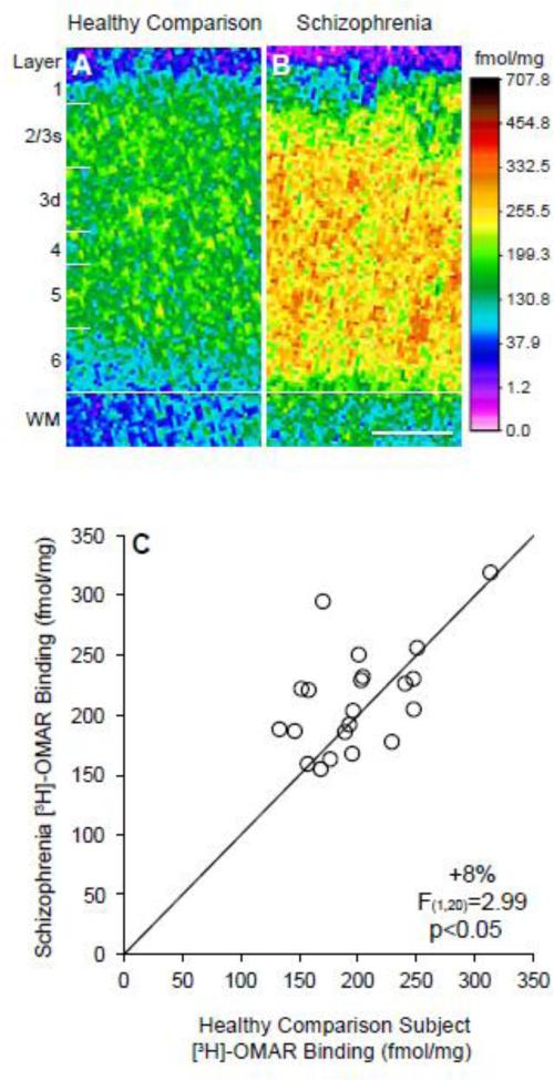
Figure 1. Receptor autoradiography for [3H]-OMAR binding in the prefrontal cortex in schizophrenia.
A-B. Pseudocolored film autoradiographs of prefrontal cortical sections processed by receptor autoradiography demonstrate higher [3H]-OMAR binding in a schizophrenia subject (B) relative to the matched comparison subject (A). Solid white line indicates the layer 6/white matter (WM) border; white distance calibration bar = 1 mm. C. Average [3H]-OMAR binding levels across gray matter of prefrontal cortical area 9 for schizophrenia subjects relative to matched healthy comparison subjects in a pair are indicated by open circles. Data points to the left of the unity line indicate higher [3H]-OMAR binding levels in the schizophrenia subject relative to the healthy comparison subject and vice versa. Mean [3H]-OMAR binding was 8% higher in schizophrenia subjects relative to matched healthy comparison subjects.

