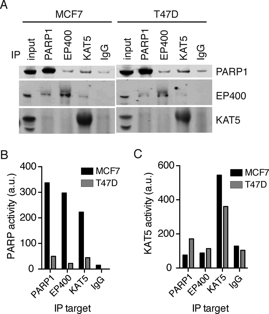
Figure 4. PARP1 forms two biochemically distinct complexes with NuA4.
A) Immunoprecipitations were performed from 1mg of total protein with the indicated antibodies. Immunoprecipitated proteins were probed for PARP1, EP400 or KAT5 by immunoblotting. The input sample contains 15ug of total protein. B) PARP1 activity, via 32P-NAD+ incorporation, and C) KAT5 activity, via in vitro acetylation of histone H4, were measured in each immunoprecipitated complex from MCF7 cell lysates (dark bars) and T47D cell lysates (light bars). Representative data from three independent experiments is shown. See also Figure S5.

