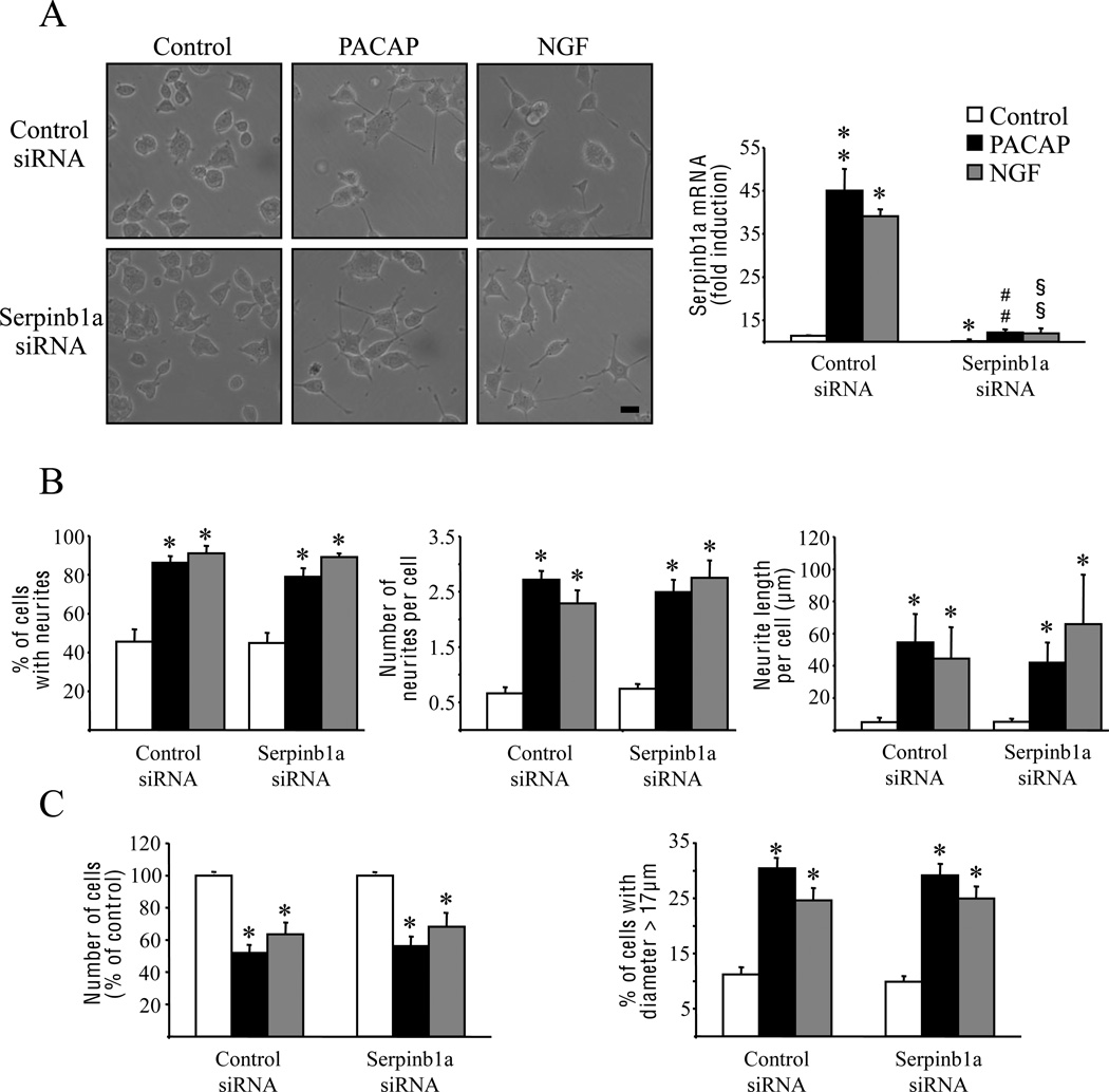
Fig. 4.
Involvement of serpinb1a in the neurotrophic effects of PACAP and NGF on PC12 cells. Cells were transfected with control siRNA or siRNA directed against serpinb1a mRNA, and were treated with PACAP (10−7 M) or NGF (100 ng/ml) 2 days later. (A) Effect of anti-serpinb1a siRNA on cell differentiation (photomicrographs) and serpinb1a mRNA expression after treatment with PACAP or NGF for 24 h and 6 h, respectively. Quantitative PCR results were corrected using the GAPDH signal as internal control and expressed as the mean fold induction (± S.E.M.) of serpinb1a mRNA level compared to untreated cells transfected with control siRNA. (B) Neuritogenesis assessment by measuring the percentage of cells with neurites, the number of neurites per cell, and the overall neurite outgrowth after 48 h of treatment with PACAP or NGF in the presence of control siRNA or anti-serpinb1a siRNA. (C) Quantification of cell proliferation and cell size by counting the number of cells and the percentage of cells with a diameter greater than 17 µm after 48 hours of treatment with PACAP or NGF in the presence of control siRNA or anti-serpinb1a siRNA. * p<0.05 and ** p<0.01 versus control in the presence of control siRNA; ## p<0.01 versus PACAP in the presence of control siRNA; §§ p<0.01 versus NGF in the presence of control siRNA. Scale bar = 15 µm.

