Figure 4.

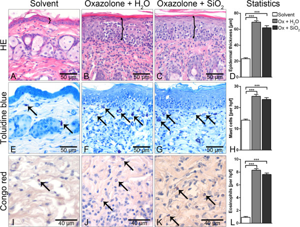
Histologic and morphometric analyses of lesional skin. Hematoxylin and eosin (HE) staining and measurement of epidermal thickness indicated by brackets (top panel) failed to reveal inflammation-associated epidermal thickening in solvent controls (A) but showed marked thickening of the epidermis and intraepidermal immune cell exocytosis in oxazolone (Ox)-treated mice without differences between the vehicle control and AHAPS-SiO2-NP treatments (B-D). Increased infiltrations with mast cells (toluidine blue, central horizontal panel: E-H) and eosinophils (Congo red, bottom panel: I-L, arrows) were very similar between the two Ox-treated groups. Only a few mast cells and eosinophils were observed in acetone solvent controls (E, I). Images represent typical samples. Quantification included all samples in panels D, H, and L. Data are presented as mean ± SEM; Ox-treated groups: n = 5; solvent-treated group: n = 3; ***p < 0.001.
