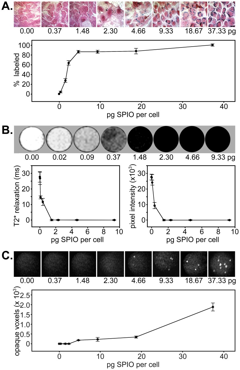
Figure 1. In vitro detection of SPIO nanoparticle-labeled muscle progenitor cells by histology, MRI, and μCT.
A) Cells incubated with increasing concentrations of SPIO particles and PLL were fixed and stained with Prussian blue and pararosaniline to identify iron-labeled cells. Slides were imaged using brightfield microscopy. The graph shows the percentage of SPIO-labeled cells at each concentration (mean ± standard error of the mean) as determined by analysis from three blinded observers. B) MRI of SPIO-labeled cell standards incorporated within fibrin sealant in micro-centrifuge tubes. The graphs display T2* relaxation time versus SPIO concentration (left) and average pixel intensity versus SPIO concentration (right) as determined from mean values generated from three different labeling experiments. C) μCT images of the same SPIO-labeled cell standards incorporated within fibrin sealant as depicted in B). The graph displays the number of opaque voxels versus SPIO concentration as determined from values generated from three different labeling experiments. Data represents mean ± standard error of the mean for each.

