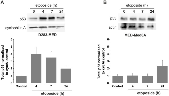
Figure 2. p53 activation is impaired in MEB-Med8A cells.
(A) D283-MED cells were treated with [20 µM] etoposide for indicated times and the p53 protein levels were measured by western blot. (B) MEB-Med8A cells were treated with [20 µM] etoposide for indicated times and p53 protein levels were measured by western blot. 2 gels from independent experiments were quantified by densitometry analysis (AQM Advance 6 imaging software, Kinetic Imaging Ltd). The plot shown is the result of the quantification relative to cyclophilin A levels and normalised to t0 untreated control ± sd for each cell line.

