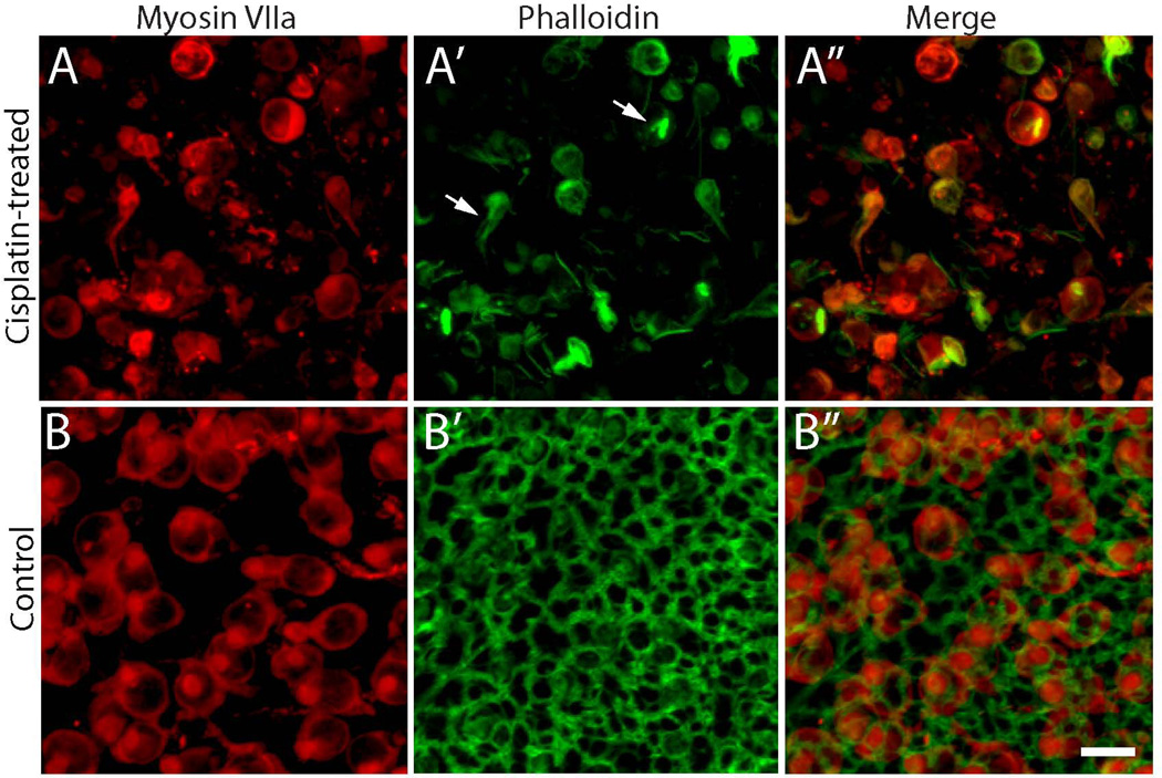
Figure 2. Cisplatin treatment disrupts the actin cytoskeleton of the utricular sensory epithelium.
Utricles were cultured for 24 hr in 10 µM cisplatin and then maintained for an additional four days in cisplatin-free medium. Immunoreactivity for myosin VIIa (red) revealed fewer hair cells in cisplatin-treated specimens (A) vs. controls (B). Labeling with phalloidin (green) and imaging with confocal microscopy indicated that cell-cell junctions were severely disrupted in cisplatin-treated utricles (A’), but appeared to be intact in control utricles (B’). Notably, some filamentous actin remained within surviving hair cells after cisplatin treatment (arrows, A’). Scale bar = 10 µm.

