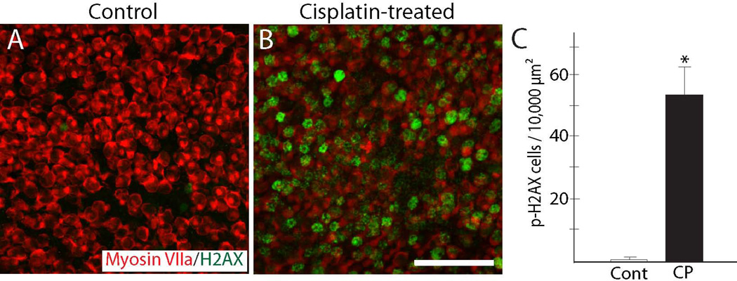
Figure 3. Evidence for DNA damage following cisplatin treatment.
Utricles were cultured for 24 hr in 10 µM cisplatin and then rinsed and maintained for an additional 48 hr in cisplatin-free medium. Control specimens were maintained in parallel (3 days, total), but did not receive cisplatin. After fixation, specimens were processed for immunohistochemical detection of p-H2AX (green), which identifies cell nuclei that contain double-stranded DNA lesions. Very few p-H2AX-labeled cells were observed in control utricles (A). In contrast, utricles that had received cisplatin treatment contained numerous cells that displayed evidence of DNA damage (B, C, *p<0.0005, t-test). CP=cisplatin-treated. Red: Myosin VIIa. Scale bar=50 µm.

